The concept of Microfocus X-ray is considered a highly precise imaging technique applied in current Non-Destructive Testing (NDT). The technology is utilized in industries in which even a minor defect will cause malfunctions, such as the manufacturing of electronics, aerospace products, automotive equipment, and semiconductors.
Unlike traditional X-rays that produce relatively large focal spots, microfocus X-rays generate a much smaller area of focus. This feature results in better image resolution compared to traditional X-ray technology, which makes the utilization of such a technique extremely important in advanced inspection cases, including industrial CT scanning involving both 2D and 3D imagery.
Microfocus X-ray techniques are especially significant when it comes to developing advanced inspection systems such as Industrial CT scanners created at XRAY LAB.
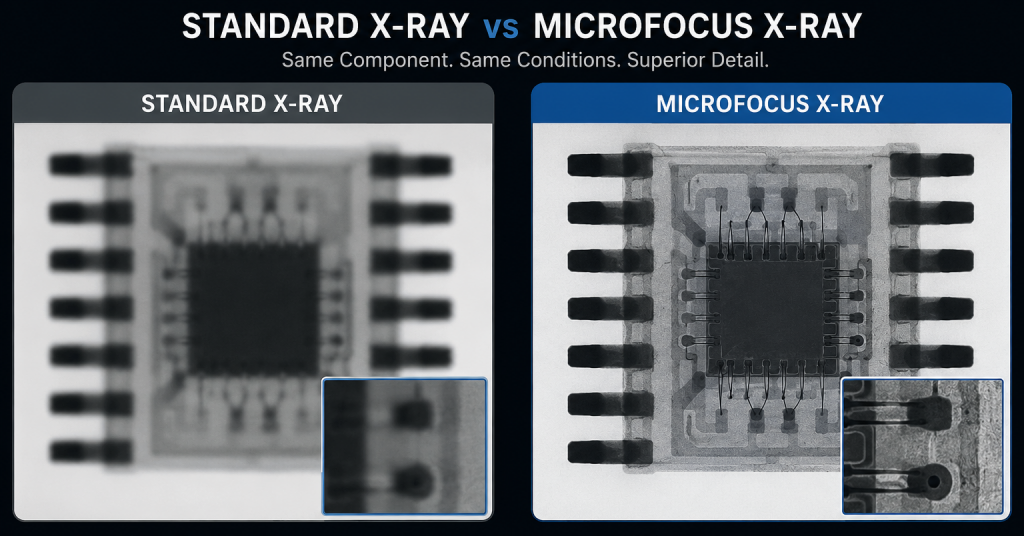
Side-by-side comparison of standard X-ray and microfocus X-ray imaging on the same electronic component, demonstrating how reduced focal spot size in microfocus systems significantly enhances image sharpness and reveals internal micro-defects with higher clarity.
What Is Microfocus X-Ray Technology?
The microfocus X-ray technology is the process whereby the electrons are concentrated on a very small area on the target in the X-ray tube to produce the focal spot, which is in micrometers. It is a very small focal spot compared to that of a standard X-ray tube.
Read the blog for Difference between Industrial X-Ray and CT scan more detailed analysis.
The basic working process includes:
- Acceleration of electrons inside the X-ray tube
- Impact of electrons on a metal target (anode)
- Generation of X-rays from a very small focal region
- Detection of transmitted radiation to create an image
The very small area of radiation source results to high degree of precision of the produced X-ray beams with minimal geometry distortion, hence enabling high resolution imaging of internal structure.
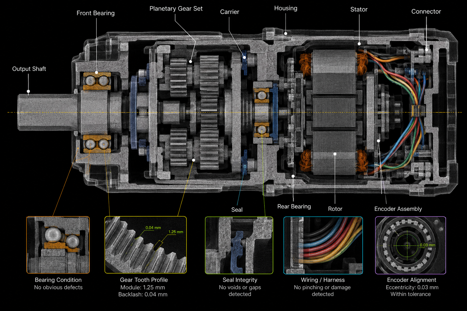
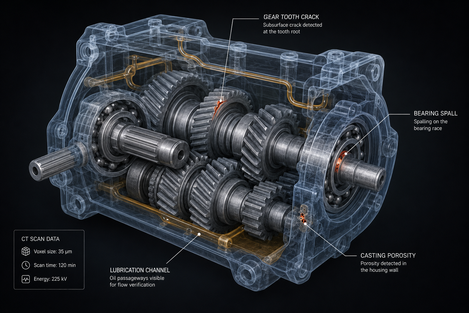
This phenomenon is particularly important in industrial CT, where several images are taken at various angles and then reassembled into three dimensional models for further analysis.
Focal Spot Size and Image Sharpness
The focal spot size is one of the most important parameters that affect image quality in X-ray systems.
A focal spot is the physical location where the electron beam impacts the anode to produce X-rays. Microfocus X-ray tubes have very tiny focal spots, which can be expressed in micrometers (µm).
Impact on image quality
Image sharpness is strongly affected by geometric unsharpness:
- Smaller focal spot → sharper edges and higher detail visibility
- Larger focal spot → increased blur and reduced resolution
This becomes crucial in the examination of finer details, like solder bonds, cracks, or voids within dense objects.
For industrial computer tomography scanners, as utilized in the XRAY LAB inspection processes, having a small focal spot becomes important in obtaining a high-quality image of the geometry inside.
Why Microfocus X-Ray Technology Is Important in Industry?
As manufacturing continues to move toward miniaturization and complex assemblies, traditional inspection methods are no longer sufficient to detect internal defects.
Microfocus X-ray technology is widely used in industries such as:
- Electronics manufacturing: Inspection of PCBs, BGA components, and micro solder joints
- Semiconductor industry: Detection of bonding defects and internal voids
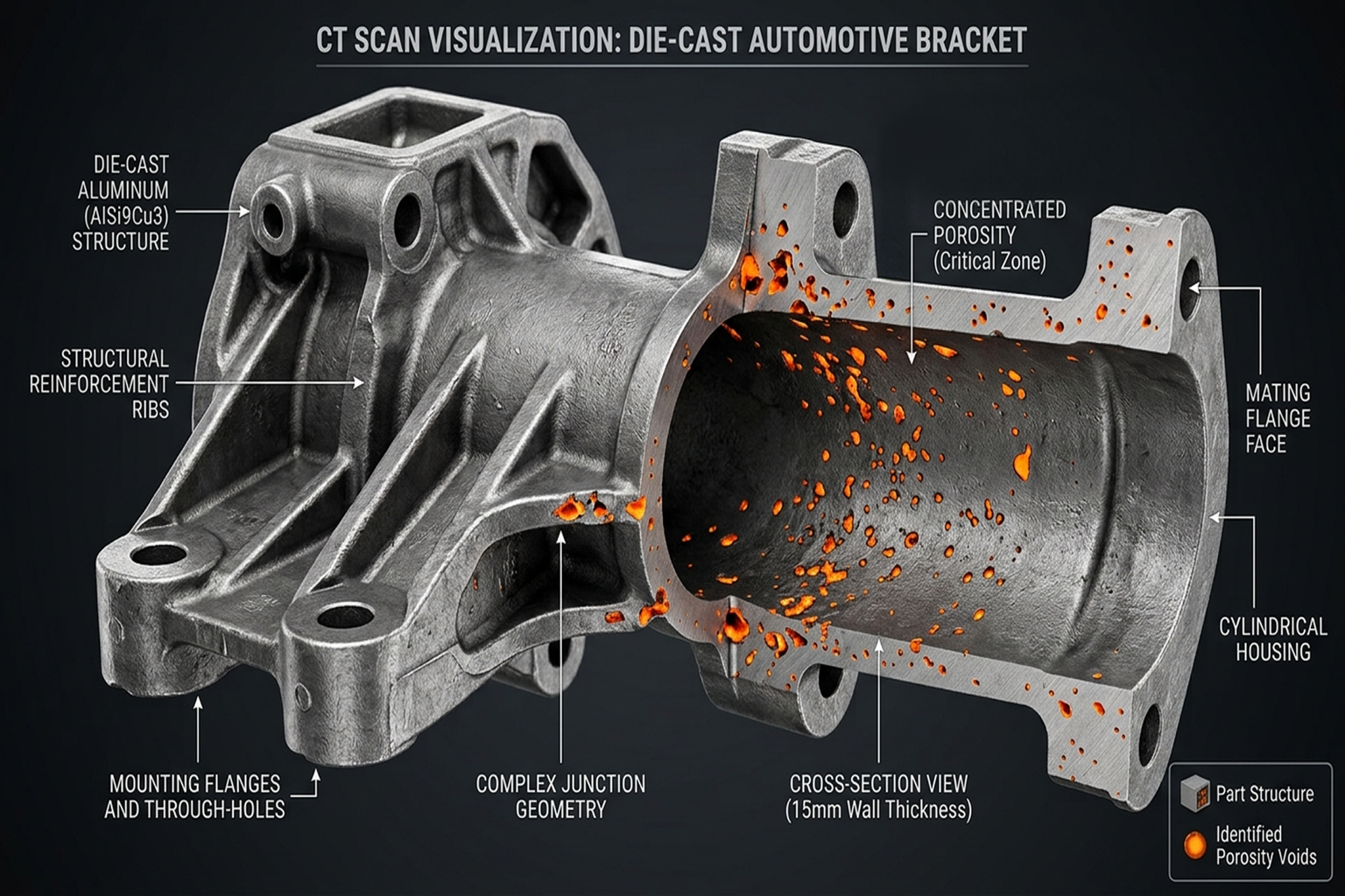
- Automotive engineering: Evaluation of lightweight and safety-critical components
- Aerospace applications: Inspection of high-precision structural parts
Microfocus technology makes possible high-resolution CT scanners used in high-level manufacturing applications by allowing precise 3D images of internal components to be created, as done at XRAY LAB.
Relationship Between Focal Spot, Magnification, and Resolution
The performance of a microfocus X-ray system depends on the interaction of several technical parameters.
1. Focal Spot Size
A smaller focal spot reduces image blur and improves spatial resolution.
2. Magnification Geometry
Magnification is controlled by system setup:
- Object closer to the source → higher magnification
- Detector positioning affects image scaling
However, magnification alone cannot improve image quality unless the focal spot is sufficiently small.
3. Spatial Resolution
Spatial resolution defines the smallest visible feature in an image and depends on:
- Focal spot size
- Detector pixel size
- System geometry and magnification
Key Insight
The high resolution in imaging comes from the use of small focal spot size together with the proper geometry and detector properties. This is the concept utilized in sophisticated CT testing machines such as the XRAY LAB machines.
Industrial Relevance of Microfocus X-Ray Systems
The use of microfocus X-rays is imperative in quality control and failure analysis due to its ability to facilitate:
Non-destructive inspection inside the component
Identification of tiny defects within the component
Imaging of complicated assemblies in great detail
Three-dimensional reconstruction of scanned parts in CT systems
With the advent of today’s high precision manufacturing environment, which requires CT-based inspection, XRAY LAB employs all the above properties for defect detection purposes.
Conclusion
Another important breakthrough in industrial inspection is microfocus x-rays, which offer very high resolution in the inspections thanks to the creation of a very small focal spot. Microfocus x-ray technology will remain relevant as industries require increased precision in their work on more complex parts.
In advanced inspection systems, including industrial CT solutions used in XRAY LAB workflows, microfocus technology is essential for achieving accurate internal visualization and ensuring product integrity across critical applications.
Frequently Asked Questions
What is microfocus X-ray technology?
Microfocus X-ray technology uses a very small focal spot to produce high-resolution images for non-destructive internal inspection of components.
Why is focal spot size important in X-ray imaging?
Smaller focal spots reduce image blur and improve sharpness, allowing detection of very fine internal defects.
What industries use microfocus X-ray systems?
Electronics, semiconductor, automotive, aerospace, and medical industries commonly use microfocus X-ray and CT systems.
How does microfocus technology improve CT scanning?
It enhances spatial resolution and enables more accurate 3D reconstruction of internal structures.
How is XRAY LAB related to microfocus X-ray technology?
XRAY LAB integrates microfocus X-ray principles into industrial CT systems to achieve high-resolution internal inspection and defect analysis.